Myocardial tissue characterization by combining late gadolinium enhancement imaging and percent edema mapping

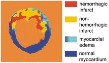 Assessing the extent of ischemic and reperfusion-associated myocardial injuries remains challenging with current magnetic resonance imaging (MRI) techniques. Therefore, the aim of this study was to develop a tissue characterization mapping (TCM) technique by combining late gadolinium enhancement (LGE) with the novel percent edema mapping (PEM) approach to enable the classification of tissue voxels as healthy, myocardial edema (ME), necrosis, myocardial hemorrhage (MH), or scar. Preliminary results suggest that the TCM method is feasible for the in vivo localization and quantification of various tissue components.
Authors: Pal Suranyi, Gabriel A. Elgavish, U. Joseph Schoepf, Balazs Ruzsics, Pal Kiss, Marly van Assen, Brian E. Jacobs, Brigitta C. Brott, Ada Elgavish and Akos Varga-Szemes